Science of Fluo Diving

Science of Fluo Diving

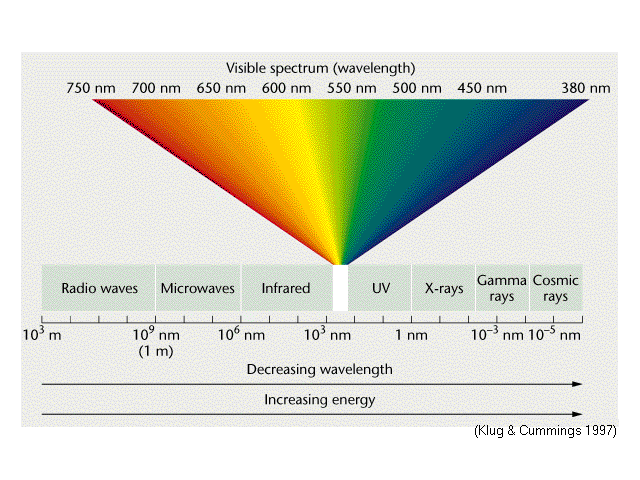
We must start our discussion with a look at the electromagnetic spectrum (EM), see the figure above. The visible portion of the EM spectrum is a very narrow band of wavelengths from about 400 to 700 nanometers (nm or 1×10⁻⁹ meters). The area we are most concerned with is what is called near ultraviolet (UV) or “actinic” light. This is light whose peak intensity is centered around a wavelength at approximately 420 nm. All radiative energy has an associated wavelength (λ) and frequency (f). One is a function of the other: f=C/λ and λ=C/f, where C is the speed of light (3×10⁸ meters per second, or m/s).


Read more

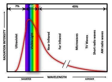
Above is a graph representing the output of the sun (solar irradiance). Note where the peak amplitude is – right there at the actinic band of wavelengths.

As a side note, the numbers at the top of the figure represent the percentages of energy emitted by the sun at various wavelengths. About 7% is in the UV range, 44% is visible light and the remaining 49% is infrared and beyond. That last 49% is felt by you as warmth.


Explore More
-->

The Physics of Light

Wes must start our discussion with a look at the electromagnetic spectrum (EM), see the figure above.

The visible portion of the EM spectrum is a very narrow band of wavelengths from about 400 to 700 nanometers (nm or 1×10⁻⁹meters). The area we are most concerned with is what is called near ultra violet (UV) or “actinic” light. This is light whose peak intensity is centered around a wavelength at approximately 420 nm.  All radiative energy has an associated wavelength (λ) and and frequency (f).

Read more

The Science of Fluorescence

The Science Behind Light Fluorescence in Dive Photography

Fluorescence is the emission of light by a substance that has absorbed light or other electromagnetic radiation. It is a form of luminescence. In most cases, the emitted light has a longer wavelength, and therefore lower energy, than the absorbed radiation. Fluorescence occurs when an orbital electron of a molecule, atom or nano-structure relaxes back to its ground state, thereby emitting a photon of light, after being excited to a higher quantum state by some type of energy. In our case, we are “hitting” an organism with higher energy light (relatively) in the near-actinic range, and lower energy light (relatively) in the green, yellow and red portion of the spectrum is being emitted. The actual color emitted is determined by how many quantum states the electron has “decayed” or relaxed back to.

Read more

Excitation Filter Discussion

Read more

Technology Discussion

Generally there are two ways of experiencing underwater fluorescence, either with invisible (ultraviolet) or with visible (usually blue) excitation light.
In the case of ultraviolet excitation light (with wavelengths <400 nm), no filters are needed for your mask and camera. You may need a “Wood’s Glass” type of excitation filter on your torch, though, in case you use a torch with white light (or a strobe), which should have lots of output in the ultraviolet range of the spectrum, such as HID (high intensity discharge) lights.
The same can be done with a white light (or strobe) and a blue excitation filter in order to obtain a blue excitation light, by the way.
However, using a white light in combination with an excitation filter that will throw away most of the torch’s light output is not as efficient as using a torch which has the desired range of wavelengths in the first place.

Read more

Discussion on Lumens

We are asked nearly every day how many lumens our blue light torches emit. Simply stated, you can’t measure a monochromatic blue light in lumens. For a VERY detailed description of why that is,- ”Lumens” is a measure of how much light the human eye will perceive, weighted according to the specific sensitivity of the human eye to different wavelengths of light. The light of our blue light torches however is not meant to eventually reach the human eye, on the contrary, it is meant to be filtered out by the barrier mask and camera filters. Instead, it is meant to pump energy into the fluorescent pigments of the underwater organisms of interest, so the most appropriate measure for our torches is the light energy or “radiant flux” they emit. What reaches the human eye (or the camera) is the fluorescent light emitted by the fluorescent pigments, whose efficiency in doing so varies greatly depending on the organism (and the pigment) in question.

Read more

Blue Light vs UV Light: What’s the Difference?

What Is UV Light?

UV light, or ultraviolet light, is a type of electromagnetic radiation. UV wavelengths are shorter than visible light, so we cannot see them with the naked eye. While the sun produces UV light alongside visible light, ~95% of all UV rays from the sun get reflected off the water’s surface.

Once divers submerge themselves over just a couple of feet deep, no UV light is present, which is why coral reefs don’t regularly glow.

Certain materials and specimens absorb UV light and reflect it back as a fluorescent glow.

Read more

Why We Use Dichroic Filters

We get technical questions everyday about the physics of fluo diving, why we design our gear the way we do and what the implication of those designs are. Follow the link below for a detailed discussion of what dichroic filters are why we use them and why you should never buy a torch without one. Dichroic filters are a type of interference filter. They have multi-layer thin-film coatings deposited on a substrate using high vacuum deposition techniques. A high voltage electron “gun” is used to vaporize various metal oxides such as aluminum, titanium, gold, silver, silica, magnesium, zirconium, chromium and various others which settle on the substrate. Each film layer is approximately one one-thousandth of a millimeter thick. The film coating typically consists of between 20 and 50 separate layers. The substrate or base material is a special type of glass or sometimes polycarbonate that has low thermal expansion properties. Dichroic filters are expensive to produce and the equipment required to produce them is very expensive.

Read more

Barrier Filter Discussion

Barrier filters are usually made from acrylic but sometimes they are also made from glass, mostly for camera filters.
The advantage of acrylic is that it is easier to cut into arbitrary shapes, and that there is less risk of injury from breaking, whereas the disadvantage is that this material is more prone to scratching. However, this latter problem is alleviated by water, which usually fills the scratches under water and renders them imperceptible.
Beware of mixing filters and light sources from varying manufacturers. Your results can vary depending on the precise spectral properties of the filter material used, the images you will see or obtain may differ significantly from those made with “spectrally matched” components.
The technology of barrier filters has come a long way over the last few years. Below is an image of how it used to be done. Image courtesy: Larry Cohen.

Read more

Spectrography

Spectroscopy is the study of the interaction between matter and radiated energy. Historically, spectroscopy originated through the study of visible light dispersed according to its wavelength, e.g., by a prism. Later the concept was expanded greatly to comprise any interaction with radiative energy as a function of its wavelength or frequency.

Spectroscopic data is often represented by a spectrum, a plot of the response of interest (amplitude) as a function of wavelength or frequency. This is often referred to as spectral power distribution (SPD).

Read more

Biology of Why Marine Life Fluoresces

It is not well understood why it is that some corals and other sea creatures fluoresce. What is known is that some marine organisms (such as corals, sponges, anemones, jellyfish, clams, nudibranchs, shrimp, crabs, worms, and fish) produce proteins that react to light causing this effect. The curious reader is encouraged to do a web search as there are several detailed peer-reviewed scientific papers on this topic and about the scientific and medical implications of this phenomenon (see e.g. Green Fluorescent Protein (GFP) for an introduction).


Read more


Additional Links

The following link is to archival sites formally operated by Steffen Beyer. Steffen was the co-creator of firedivegear.com/old and operated a couple of parallel fluo information sites. Much of the information here is quite dated but there are still some good references here.  Sadly, due to personal issues, he is no longer able to support the links below so there must be a small disclaimer that some of the sublinks will be broken.

http://guest.engelschall.com/~sb/fluolinks/

Read more


If you have any questions, comments or suggestions – PLEASE don’t hesitate to contact us.

Translate »